Andreia Maia Oliveira (CERN & University of Bern)
An advantageous radiation modality for treating cancer is hadron therapy, which at present uses protons and carbon ions. However, treatment with hadrons requires high spatial and dosimetric accuracy in order to achieve optimal dose delivery, which is in turn guaranteed by proper quality assurance (QA) procedures and tools.
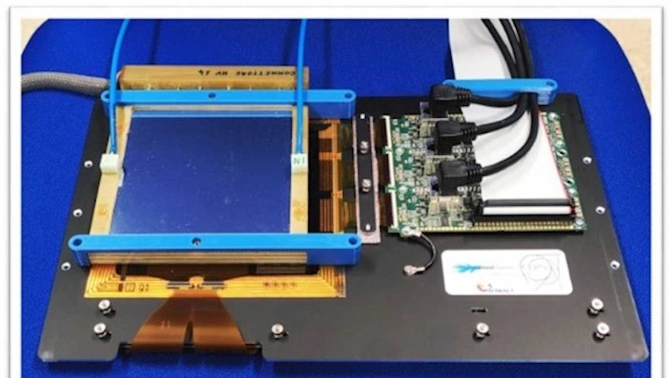
For this purpose, a promising tool is the LaGEMPix detector that combines a triple Gas Electron Multiplier (GEM) with a highly pixelated readout. A first prototype of the LaGEMPix based on a matrix of organic photodiodes (OPD) fabricated on top of an oxide-based thin-film transistor (TFT) backplane with an active area of 60 x 80 mm2 and pixels of 126 x 126 μm2 has been successfully built and tested with X-rays. The spatial resolution obtained with the LaGEMPix of 2.4 mm using X-Rays is insufficient. An upgraded version of the LaGEMPix has been developed and tested. However, the desired target of submillimetre spatial resolution is hardly reachable for a compact system with an optical readout without the introduction of lenses in the set-up.
A new prototype of the LaGEMPix was built. To further increase the spatial resolution, OPD frontplane present in the LaGEMPix with optical readout was eliminated, leaving a TFT-only electronic readout. We assembled and tested a charged readout GEM-based detector based on a matrix of thin-film transistors using low-energy X-rays, protons and carbon ions. This detector with TFT-based readout can measure secondary electrons produced by the GEMs with submillimetre resolution, and hence it represents an important step towards the development of a 20x20 cm2 detector to cover the typical maximum clinical radiation field size used in hadron therapy.


